Students at Cranfield University have designed computer models that use computer vision and artificial intelligence (AI) to analyse chest X-ray imagery.
The AI is able to detect anomalies in an X-ray, classifying which are positive for pneumonia–a common symptom of COVID-19. A second model is then used to diagnose if the pneumonia is caused by the COVID-19 virus.
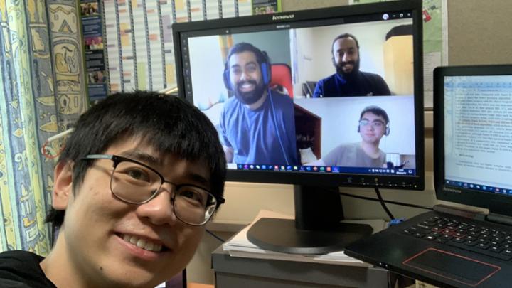
The MSc progamme students, specialising in Computer and Machine Vision, worked on this project remotely as they were impacted by the lockdown. Yet, they used the video conferencing and IT facilities provided by the University to access the necessary computational resources.
The lack of X-ray imagery in the public domain containing COVID-19 details was a challenge. However, the teams were able to build detailed information from various sources.
The AI model
The groups employed conventional machine learning algorithms as wells as deep learning frameworks, a machine learning technique that teaches computers to learn by example.
The AI model employed in this project was able to predict results with great accuracy. However, the research teams believe they are able to further develop new algorithms to produce even more robust and reliable results.
The teams are led by Zeeshan Rana, Lecturer in Computational Engineering at Cranfield University.
He is now exploring collaboration opportunities with medical authorities or industry to develop the project to the next level, using more advanced AI algorithms and CT (computed tomography) scans to show greater detail and accuracy in the results.